
How is a fatty tumor in dogs checked? A veterinarian examines the lump, then takes a few cells with a thin needle, called a fine needle aspirate, and looks at them under a microscope. If the lump needs to come out, surgery under general anesthesia removes it along with a margin of surrounding tissue, and the removed lump can be sent to a lab for a final answer. Touch alone cannot tell a harmless lump from a dangerous one, so the check comes first.
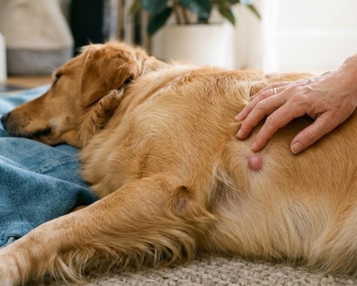
Finding a new bump while you are petting your dog is a stomach-dropping moment, and the first thing you do is press on it and wonder. That reaction is normal. A lump under a dog’s skin can easily turn out to be a benign fat deposit, and checking one is quick enough to replace the worry with an actual answer.
This guide explains what a fatty tumor is and why it is not always alarming, why a lump cannot be judged by feel, how a vet checks it step by step, how the different kinds of lumps compare, when removal makes sense, how the surgery works, what recovery looks like, what testing cannot promise, and which signs should not wait.
What a Fatty Tumor in Dogs Is and Why It Is Not Always Alarming
A fatty tumor is a lipoma, and a dog lipoma is simply a growth made of fat cells. Cornell University’s College of Veterinary Medicine describes lipomas as among the most common benign tumors in dogs, usually slow-growing, soft and movable under the skin. They usually appear on the torso and limbs, although they can also form between muscles or, less commonly, on internal organs. Some dogs have one, and some collect several.
The cause is unknown, but the pattern is familiar. Cornell says older dogs, overweight dogs and breeds such as Labrador Retrievers are more likely to develop them. Texas A&M’s veterinary college adds that a lipoma is usually painless, which is part of why owners find one by accident and not because the dog complains.
Benign does not mean ignorable. Summit’s growth and mass removal page points out that harmless growths such as warts, skin tags, cysts and lipomas can still grow quickly and interfere with normal function. A lump in the wrong spot can bother a dog long before it becomes large.
Why You Cannot Tell a Harmless Lump by Touch
A soft, movable lump feels reassuring, and that is exactly the trap. Texas A&M states that touch alone should not be used to diagnose a lump, and Summit’s own page says owners cannot reliably separate benign growths from malignant ones by looking at them.
The reason is that very different growths share the same appearance from the outside. A fatty lump, a cyst and certain cancers can all sit under the skin as a round bump. Malignant growths, in Summit’s words, can be fast-growing, destroy nearby tissue and may spread through the body. The only way to know which one you are dealing with is to look at the cells.
That is why a new lump deserves a prompt exam even when your dog seems perfectly fine. Waiting does not give the answer. It only gives the lump time to grow, which can make a future surgery larger.
How the Vet Checks a Lump Step by Step
The check begins with a conversation and a hands-on exam. The veterinarian will want to know when you first noticed the lump, whether it has changed in size or firmness, whether there are others, and whether your dog seems bothered by it. Measuring the lump and noting its location gives a baseline for later comparison.
The fine needle aspirate
The next step is usually a fine needle aspirate. Cornell describes it as a minimally invasive test in which a small needle collects cells from the mass for microscopic examination. Summit’s page describes the same idea: a few cells are removed with a needle and examined to help identify the tumor type and plan surgery. It is a quick test done with a thin needle.
The biopsy
When the aspirate does not settle the question, or when the lump needs to come out anyway, a biopsy is the next step. A biopsy examines a piece of tissue, or the whole mass, which gives a pathologist more to work with than loose cells. Cornell notes that if the entire mass is removed, the biopsy can also be the treatment for a suspected lipoma. Summit’s pet biopsy service covers this part of the process.
Checking the rest of the dog
If a lump turns out to be an aggressive cancer, Summit’s page explains that staging follows, with X-rays of the lungs to look for secondary tumors, a check of the lymph nodes that drain the area, and an examination of the liver and abdomen. Blood tests are also run to look for underlying problems and to decide whether IV fluids will be needed during anesthesia.
Simple, Infiltrative and Malignant Lumps at a Glance
Not every fat-based lump behaves the same way. The table below summarizes the groups Cornell describes, plus the lumps that are not fatty at all.
Type of lump |
What it is |
How it behaves |
What the plan involves |
| Simple lipoma | A benign, encapsulated mass of fat cells | Slow-growing, soft and movable | Monitoring, or removal if it is in an awkward spot or growing |
| Infiltrative lipoma | A benign fatty tumor that invades muscle, bone and nerves | Grows faster and comes back more readily than a simple lipoma | Wide surgical excision, sometimes with imaging first |
| Liposarcoma | A rare malignant fat-cell tumor | Does not tend to spread far but invades nearby tissue | Wide excision, sometimes with imaging or radiation therapy |
| Other lumps | Cysts, warts, skin tags and cancers that feel similar | Varies from harmless to aggressive | Needle or tissue sampling decides the plan |
When Removal Makes Sense and When Watching Is Fine
Not every lipoma needs surgery. Cornell states that not all of them need treatment, and Texas A&M says the typical lipoma causes no trouble unless it presses on a vital structure or grows big enough to interfere with movement. A simple lipoma that is not removed should be watched for changes in size, firmness or discomfort.
Removal becomes reasonable in a few situations. Cornell mentions lipomas in places such as the armpit, where the lump can get in the way of a leg. Texas A&M says surgery is generally considered for deeper lumps, such as the thigh, or when an infiltrative tumor is suspected. Cornell also notes that some veterinarians prefer to remove a lipoma before it gets large, to reduce the risk of complications after surgery.
There is no universal rule, and the decision depends on the individual dog. Age, other health conditions and whether the dog will be under anesthesia for another reason all matter. A senior dog with a small, stable lump might be monitored, while the same lump in a young dog’s armpit might be addressed sooner. Summit’s senior pet care approach is a good place to see how age shapes those choices.
How Surgery Removes the Lump
Dog mass removal is a planned surgery, and much of the safety is in the preparation. Summit’s page describes blood tests beforehand, then general anesthesia, after which the skin around the mass is cleared. The surgeon removes the lump, closes the incision with sutures and adds a dressing where needed.
What the surgeon takes matters as much as the lump itself. Summit notes that success depends on the type of growth, how early it was caught, how accessible it is, how much surrounding tissue is removed and the surgical technique. For a simple lipoma that sits cleanly under the skin, removal can be straightforward. For an infiltrative lipoma, Cornell explains that a wide excision is usually needed so that no tumor cells are left behind, and imaging such as CT may be used for planning, with radiation therapy as an option in some cases.
The removed tissue should go to a laboratory. That step turns a presumed fatty lump into a confirmed diagnosis, and it tells you whether the surgeon got everything. For details on the procedure itself, Summit’s surgery page lists what the hospital performs.
Recovery and What Healing Looks Like
Summit says pets can be somewhat groggy for a day or two after the surgery and that the veterinarian provides post-operative care instructions. Those instructions are the plan to follow, because wound care and activity limits depend on where the incision is and how large the lump was.
One complication is worth knowing about. Cornell explains that when a large mass is removed it can leave a space, and that space can fill with fluid, a swelling called a seroma. If you notice a soft swelling near the incision after surgery, mention it to the clinic and do not try to drain it yourself.
The long-term picture for a simple lipoma is encouraging. Cornell reports an excellent prognosis after complete removal, and recurrence is rare, although a dog prone to lipomas can develop new ones elsewhere. For infiltrative lipomas and liposarcomas, complete removal is harder to achieve, which raises the risk of regrowth, but a good outcome is still possible if the tumor is fully removed.
What Testing and Surgery Cannot Promise
A needle aspirate is quick, but Texas A&M points out that it cannot tell a simple lipoma from an infiltrative one. Telling those apart takes advanced imaging or surgery. Summit’s radiology page says the hospital refers CT and MRI to outside specialists, so if your dog needs that level of imaging, the team will help you arrange it.
A confirmed lipoma also does not protect against other lumps. A dog can have a fatty tumor and a separate growth that behaves differently, so every new lump deserves its own check.
Finally, no surgery guarantees that a lump will not return. A fully removed simple lipoma rarely comes back, but the tendency to grow lipomas stays with the dog. A tumor that spreads into surrounding tissue can regrow even after careful surgery.
Lump Signs That Should Not Wait
Any new lump is worth a call. These changes make the call more urgent.
- A lump that grows noticeably from one week to the next is a change that should be checked soon.
- A lump that turns hard, red, warm or painful, or that opens and drains, needs an exam before it can become infected.
- A dog who limps or avoids moving a leg because of a lump near a joint or in the armpit may have a lump that is interfering with movement.
- A dog who loses weight, eats less or seems tired while a lump grows should be examined to see whether something else is going on.
- A lump that appears suddenly, or several new lumps in a short time, should be examined and not assumed to be fat.
For urgent problems after hours, Summit’s urgent care and emergency care page explains what the hospital can handle.
How Summit Supports Dogs With Lumps
The growth and mass removal service at Summit follows the path described above: an exam, a needle sample sent for analysis, bloodwork, anesthesia and surgery when it is needed, and written care instructions afterward. Imaging that goes beyond the hospital’s own equipment is referred out, and the team can explain when that step is worthwhile.
For dogs in Summit, Short Hills, Millburn, Livingston, Chatham and surrounding towns, the practical advantage is speed. Same-day and next-day appointments mean a lump does not have to be a week-long worry before someone looks at it.
From Lump to Answer
A fatty tumor in dogs is usually benign, but you cannot know that by touch. A fine needle aspirate gives a first answer, a biopsy confirms it, and surgery removes the lump when it is causing trouble or when the diagnosis calls for it.
If you have found a lump on your dog, Summit Dog and Cat Hospital can check it and explain the options. You can book a same-day or next-day appointment online, or call to talk it through.
Sources
Cornell University College of Veterinary Medicine explains lipomas in dogs, how they are diagnosed, when surgery is recommended and what recovery involves.
Texas A&M University College of Veterinary Medicine describes how fatty tumors are checked, what a needle aspirate can and cannot show and when removal is considered.
Frequently Asked Questions
Q: How is a fatty tumor in dogs diagnosed?
A: Your veterinarian examines the lump and usually takes a fine needle aspirate, collecting a few cells for a microscope. A biopsy may follow if the answer is unclear or the lump needs to come out. Feel alone is not reliable, so the cells get checked.
Q: Does a dog lipoma always need to be removed?
A: No. A simple lipoma that is not causing problems can be monitored for changes in size, firmness or comfort. Removal is considered when it sits where it interferes with movement, grows, or is suspected to be the infiltrative type.
Q: What does dog mass removal involve?
A: Your dog receives general anesthesia, the skin around the lump is cleared, and the surgeon removes the mass and closes the incision with sutures. The tissue can be sent to a laboratory for a final diagnosis. Your veterinarian will send written care instructions home.
Q: How long does a dog take to recover after a lump is removed?
A: A dog may be groggy for a day or two, and your veterinarian will explain wound care and activity limits for your dog. Healing depends on the size and location of the lump, so follow the instructions you are given and report any swelling near the incision.
Q: Can I book a same-day appointment at Summit for a new lump?
A: Summit offers same-day and next-day appointments, so a new lump does not have to wait. Book online or call the hospital, and the team will tell you what is open.